 Jul, 28 2026
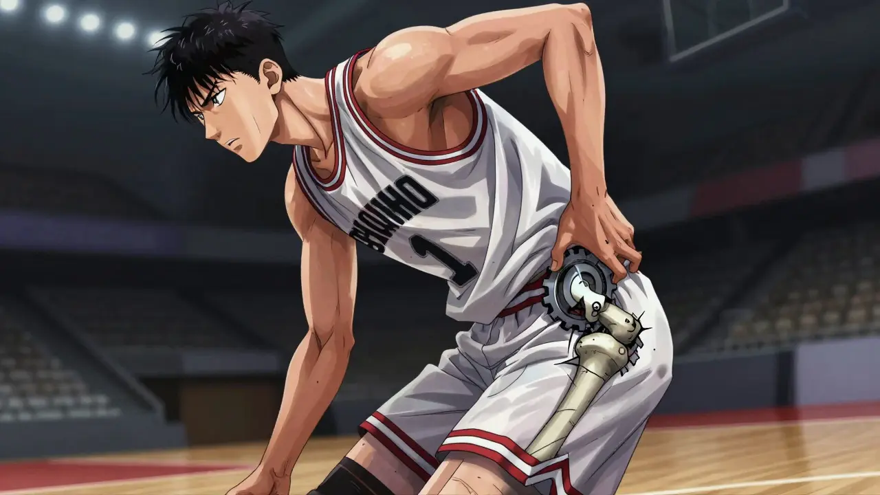
That sharp pinch in the front of your groin when you twist or squat isn't just a muscle strain. For many athletes, it’s a signal that something deeper is wrong with the hip joint itself. A hip labral tear is damage to the fibrocartilaginous rim (labrum) that lines and protects the acetabular socket of the hip joint. This injury has become one of the most common reasons for persistent hip pain in sports, particularly among those under 40 who play basketball, soccer, football, or hockey.
If you are an athlete dealing with this issue, the path to recovery can feel confusing. You might be told to rest, then referred for scans, and finally offered surgery. Understanding what is actually happening inside your hip, how doctors see it, and why some people need arthroscopy while others do not, is crucial for getting back to the game you love.
Understanding the Hip Labrum and Why It Tears
To understand the tear, you first have to understand the part that tore. The hip is a ball-and-socket joint. The "ball" is the head of the femur (thigh bone), and the "socket" is the acetabulum in your pelvis. Lining the edge of that socket is the labrum, which acts like a suction cup seal to deepen the socket and stabilize the joint.
When this seal tears, stability drops, and pain spikes. But why does it happen? The leading cause is femoroacetabular impingement (FAI), a condition where abnormal bone growth causes the bones of the hip to rub against each other during movement. Think of it like a door hinge that doesn't sit quite right; every time you open and close the door, the metal grinds against the frame. In the hip, this grinding pinches the soft labrum between the hard bones, eventually causing it to rip.
This isn't just about bad luck. Approximately 22-55% of all athletic hip pain cases involve labral pathology. If you play sports that require repetitive hip rotation-like cutting in soccer or pivoting in basketball-you are in the highest-risk demographic. The mechanics of these sports place extreme stress on that delicate cartilage rim.
The Diagnostic Challenge: Seeing What Plain X-Rays Miss
One of the biggest frustrations for athletes is the diagnostic process. You go to the doctor, they look at your hip, maybe order an X-ray, and tell you everything looks fine. But it hurts. Here is the problem: standard imaging often misses the damage.
Plain radiographs (X-rays) are the starting point, but they only show bone. They are excellent for spotting FAI, dysplasia (a shallow socket), or arthritis, but they cannot see the soft tissue of the labrum. If your X-ray is normal, it doesn't mean your hip is healthy; it just means your bones aren't broken or severely arthritic yet.
Next comes the MRI. Standard MRIs have a sensitivity of only 35-60% for detecting labral tears. That means nearly half of the time, a standard MRI will miss the tear entirely. This is why many patients get false reassurance.
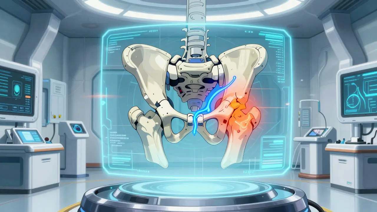
The gold standard for non-surgical diagnosis is magnetic resonance arthrography (MRA), an enhanced MRI where contrast dye is injected directly into the hip joint. The dye leaks out through any tears in the labrum, lighting them up clearly on the scan. MRA boasts a 90-95% sensitivity rate. If you have been diagnosed with a "normal hip" but still have mechanical symptoms like clicking, catching, or locking, ask your specialist about MRA. It provides the clarity needed for accurate treatment planning.
| Imaging Method | Sensitivity | Best Used For | Limitations |
|---|---|---|---|
| Plain Radiograph (X-Ray) | N/A (Soft Tissue Blind) | Assessing bone structure, FAI, and dysplasia | Cannot visualize the labrum or cartilage |
| Standard MRI | 35-60% | Ruling out other soft tissue injuries | Frequently misses partial-thickness labral tears |
| Magnetic Resonance Arthrography (MRA) | 90-95% | Detecting labral tears and cartilage defects | Invasive (requires injection); higher cost ($1,200-$1,800) |
| Hip Arthroscopy | 98% | Definitive diagnosis and simultaneous treatment | Surgical procedure with associated risks |
Conservative Treatment: Can You Avoid Surgery?
Not every tear requires a surgeon's knife. In fact, many athletes successfully manage their symptoms without going under anesthesia. The initial protocol usually involves 4-6 weeks of relative rest, avoiding activities that trigger the pinch, and using non-steroidal anti-inflammatory drugs (NSAIDs) like ibuprofen to control inflammation.
Physical therapy is the cornerstone of conservative care. However, results vary. Some studies suggest only 30-40% of athletes achieve complete resolution through therapy alone, while other clinics report success rates as high as 65%. The difference often lies in the type of therapy. Generic stretching won't cut it. You need targeted strengthening of the gluteal muscles and core stabilizers to offload pressure from the hip joint.
If pain persists, doctors may recommend corticosteroid injections. These provide temporary relief in 70-80% of cases, lasting 3-6 months. This window of relief is critical-it allows you to engage more effectively in physical therapy. Newer approaches include platelet-rich plasma (PRP) injections, which show promise in helping 55% of patients avoid surgery at the 12-month mark by promoting natural healing.
However, if conservative measures fail after 3-6 months, or if the tear is large and unstable, surgery becomes the standard option. Delaying necessary surgery can lead to further cartilage damage, increasing the risk of early-onset osteoarthritis.
Hip Arthroscopy: The Surgical Solution
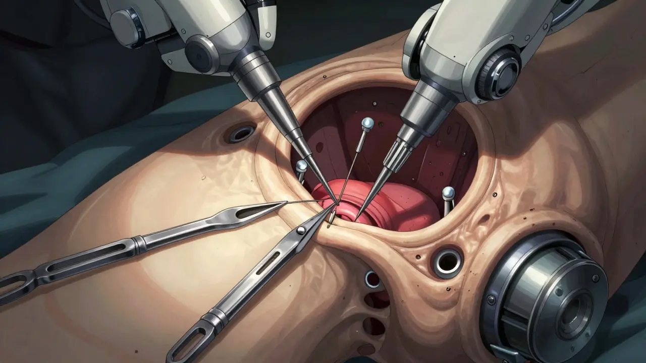
When non-surgical options run out, hip arthroscopy is the primary surgical intervention. This minimally invasive procedure involves making small incisions and inserting a camera and instruments into the hip joint. It allows surgeons to directly visualize the damage with 98% accuracy.
There are two main surgical strategies:
- Debridement: The surgeon trims away the torn, frayed edges of the labrum to smooth it out. This is less invasive but doesn't restore the original seal function of the labrum.
- Repair: The surgeon uses suture anchors to reattach the torn labrum back to the acetabular rim. This preserves the joint's stability and is generally preferred for younger, active patients.
A critical factor in surgical success is addressing underlying anatomy. If you have hip dysplasia (a shallow socket), simply repairing the labrum has a 65% failure rate at two years. In these cases, surgeons must also perform a pelvic osteotomy to correct the bone structure. Ignoring the root cause guarantees the tear will come back.
Recovery timelines depend on the procedure. Debridement typically requires 3-4 months before returning to sport, while a full repair necessitates 5-6 months. Professional athletes, like NHL player Ryan Nugent-Hopkins, have returned to elite competition within 5-6 months following rigorous rehabilitation protocols.
Return to Sport and Long-Term Outlook
Getting back to playing is the ultimate goal, but rushing it is dangerous. Rehabilitation is not passive; it is an active process divided into phases. The first six weeks focus on protection and restoring range of motion. Weeks 7-12 involve progressive strengthening. Only after week 13 do athletes begin sport-specific training.
Before clearing you for return, your medical team should verify specific milestones: 90% quadriceps strength symmetry compared to your uninjured leg, and pain-free hip internal rotation to at least 30 degrees. Meeting these criteria significantly reduces the risk of re-injury.
Long-term outcomes are generally positive. Competitive athletes demonstrate an 85-90% return-to-play rate after arthroscopic intervention. However, age matters. For athletes over 35, this rate drops to 70-75%, often due to concurrent degenerative changes in the joint. Sports requiring extreme ranges of motion, such as ballet or gymnastics, carry a 25% higher complication rate post-surgery.
The good news is that treating the tear now prevents worse problems later. Untreated labral tears are associated with a 4.5 times higher risk of developing hip osteoarthritis within 10 years. By addressing the issue through proper imaging and timely intervention, you protect your hip for decades to come.
How long does it take to recover from hip arthroscopy?
Recovery time depends on the type of surgery performed. For debridement (trimming the tissue), athletes typically return to sport in 3-4 months. For labral repair (reattaching the tissue), the timeline extends to 5-6 months. Full rehabilitation involves four phases spanning up to 26 weeks, focusing initially on protection and gradually progressing to sport-specific movements.
Is MRA better than a standard MRI for diagnosing hip labral tears?
Yes, MRA is significantly more accurate. Standard MRIs have a sensitivity of only 35-60% for labral tears, meaning they miss many cases. Magnetic resonance arthrography (MRA) uses contrast dye injected into the joint to highlight tears, achieving 90-95% sensitivity. It is considered the diagnostic test of choice for suspected labral pathology.
Can physical therapy cure a hip labral tear?
Physical therapy can manage symptoms effectively for many patients, with success rates ranging from 30% to 65% depending on the severity of the tear and the quality of the rehab program. It focuses on strengthening surrounding muscles to stabilize the joint. However, if the tear is large or accompanied by structural issues like FAI, therapy alone may not resolve the mechanical symptoms, and surgery might be necessary.
What is femoroacetabular impingement (FAI)?
Femoroacetabular impingement (FAI) is a condition where abnormal bone growth on the femoral head or acetabulum causes the bones to rub against each other during hip movement. This friction pinches the labrum, leading to tears. It is the most common causative factor for hip labral tears in athletes.
What are the risks of leaving a hip labral tear untreated?
Leaving a labral tear untreated increases the risk of progressive joint damage. Studies show that untreated tears are associated with a 4.5 times higher risk of developing hip osteoarthritis within 10 years. Additionally, ongoing pain and mechanical symptoms can limit athletic performance and daily activities.